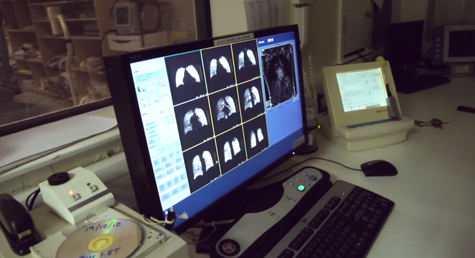
For the final Royal Institution Advent film, I travelled to the University of Sheffield MRI Unit at the Royal Hallamshire Hospital, to look at how a very strange element is being used in a pioneering MRI technique to image living lungs. http://youtu.be/dmPmHSVqfZE
The film is presented by this year's Christmas Lecturer, Dr Peter Wothers (University of Cambridge) who takes part in the research programme by having his own lungs scanned. Conventional MRI is usually pretty poor at imaging areas such as the lungs, which have very little fatty tissue and water (MRI scanners essentially detect radio frequencies given off by protons in Hydrogen nuclei) - and so this novel technique involves the inhalation of hyper-polarised Xenon to image the ventilated lung. Xenon is an inert gas so is relatively safe to inhale, although it does have some unusual effects on the human body, especially on the voice - it's also a mild anaesthetic - so watch the film to see how it affects Peter!
Xenon Lungs
As the Xenon is only present within the respiratory system, signal is only detected within ventilated areas - areas in which Xenon is not present appear black on the resulting image. This therefore allows medical professionals to identify damaged or obstructed areas of the lung which may be poorly ventilated or not at all, providing a novel method of efficiently and non-invasively examining the lungs of a living patient.

The research is being conducted by Dr Jim Wild and his research assistant Helen Marshall (both featured in the film) at the University of Sheffield and is funded by the EPSRC. More information on this technique can be found here.
The films forms part of a series of 24, released daily in the Ri Advent Calendar here. The films are also available on YouTube and on the Ri Channel.
